Christian Ledig
Quick Links
 @LedigChr


Available datasets:
Available software packages:
- Multi-Atlas Label Propagation with EM-refinement (MALPEM)
- Patch-based Evaluation of Image Segmentation (PEIS)
- Latent Tree (LT) Generative Model
MALPEM-ADNI: Features, binary masks, segmentations for 5074 ADNI subjects
We employed a recently validated method (MALPEM) for robust cross-sectional and longitudinal segmentation of MR brain images from the Alzheimer's Disease Neuroimaging Initiative (ADNI) cohort. Specifically, we segmented 5074 MR brain images into 138 anatomical regions and extracted time-point specific structural volumes and volume change during follow-up intervals of 12 or 24 months.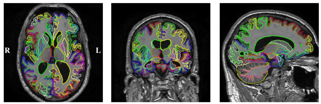 The following is a summary of the publicly available dataset. For more details please refer to the [doi] and the README of the current GIN repository.
Processed Images from the ADNI cohort
- List of all 5074 images (baseline and follow up images, incl. VISCODE)
- List of 1069 baseline images (incl. disease label) used for analysis
- List of 802 baseline and m12 follow up image pairs (incl. disease label) used for analysis
- List of 532 baseline and m24 follow up image pairs (incl. disease label) used for analysis
- All extracted cross-sectional (structural volumes, asymmetry) and longitudinal (volume change rate) features and selected clinical information (e.g. disease labels). Note: Not all of those features have been used in the manuscript. Please, refer to the paper for details.
- 5074 cross-sectional structural segmentations in 138 distinct anatomical regions (calculated with MALPEM)
- 5074 binary brain masks (calculated with pincram). Masks have been quality checked (baseline: visual, followup: automatic)
- Lookup table for all segmented 138 brain structures
C. Ledig, A. Schuh, R. Guerrero, R. A. Heckemann and D. Rueckert, “Structural brain imaging in Alzheimer's disease and mild cognitive impairment: biomarker analysis and shared morphometry database”, Scientific Reports, 8, 2018. [doi] [pdf] [bib]
(please cite for dataset)
C. Ledig, A. Schuh, R. Guerrero, R. A. Heckemann and D. Rueckert, “Dataset - Structural brain imaging in Alzheimer's disease and mild cognitive impairment: biomarker analysis and shared morphometry database”, G-Node, 2018. [dataset] [bib]
Multi-Atlas Label Propagation with EM-refinement (MALPEM)
Software package to perform a whole-brain segmentation of a T1-weighted magnetic resonance brain image. The essential modules of MALPEM are: N4 bias correction, pincram brain extraction, label propagation (ireg), label fusion (Gaussian weighted local label fusion), label refinement (EM algorithm).MALPEM should run on any 64-bit Linux machine or on MacOS/Windows using a Virtual machine. Please see 'README' file provided in the package for more information.
[Download installer: MALPEM v1.2 (20 kB)]
[Source available on GitHub]
[README]
[Example PDF report]
 Description:
To run MALPEM download the above package and run './malpem_installer/malpem-install' for installation. For further instructions on how to install and run MALPEM see provided 'README' file. MALPEM will calculate a binary brain mask, a structural segmentation (138 structures) and a PDF report.
Description:
To run MALPEM download the above package and run './malpem_installer/malpem-install' for installation. For further instructions on how to install and run MALPEM see provided 'README' file. MALPEM will calculate a binary brain mask, a structural segmentation (138 structures) and a PDF report.The methodology is described in the following publications:
(please cite for MALPEM framework and structural segmentation)
C. Ledig, R. A. Heckemann, A. Hammers, J. C. Lopez, V. F. J. Newcombe, A. Makropoulos, J. Loejoenen, D. Menon and D. Rueckert, “Robust whole-brain segmentation: application to traumatic brain injury,” Medical Image Analysis, 21(1), pp. 40-58, 2015. [pdf] [doi] [bib]
(please cite for brain extraction)
R. Heckemann, C. Ledig, K. R. Gray, P. Aljabar, D. Rueckert, J. V. Hajnal, and A. Hammers, “Brain extraction using label propagation and group agreement: pincram,” PLoS ONE, 10(7), pp. e0129211, 2015. [doi][pdf][bib]
(Additional related publications)
C. Ledig, R. A. Heckemann, P. Aljabar, R. Wolz, J. V. Hajnal, A. Hammers, and D. Rueckert, “Segmentation of MRI brain scans using MALP-EM,” MICCAI 2012 Grand Challenge and Workshop on Multi-Atlas Labeling, pp. 79-82, 2012. [pdf] [bib]
C. Ledig, R. Wolz, P. Aljabar, J. Loetjoenen, R. A. Heckemann, A. Hammers, and D. Rueckert, “Multi-class brain segmentation using atlas propagation and EM-based refinement,” Proceedings of ISBI 2012, pp. 896-899, 2012. [pdf] [bib] [doi]
Patch-based Evaluation of Image Segmentation (PEIS)
Here you can download the source code of PEIS that was implemented in C++ within the image registration toolkit (IRTK), which is provided as precompiled libraries.Please note that PEIS works on segmentations in NIfTI format (.nii, .nii.gz). Segmentations stored in other file formats can be converted to NIfTI format using for example Convert3D.
[Download PEIS]
Contains:
- Ubuntu 13.04, 64 bit: precompiled libraries/binary
- Windows 7, 64 bit: precompiled libraries/binary
- MacOSX Mavericks 10.9.3, 64 bit: precompiled libraries/binary
- MacOSX Snow Leopard 10.6.8, 64 bit: binary only
This software package implements the following paper:
C. Ledig, W. Shi, W. Bai, and D. Rueckert, “Patch-based evaluation of image segmentation,” CVPR, pp. 3065-3072, 2014. [bib] [pdf]
Latent Tree (LT) Generative Model
The Latent Tree Generative Model was implemented in Matlab by Sebastian Kaltwang and is available on GitHub.[Latent Tree on GitHub]
The Latent Tree (LT) model was first proposed in:
S. Kaltwang, S. Todorovic, M. Pantic, “Latent Trees for Estimating Intensity of Facial Action Units” IEEE Conference on Computer Vision and Pattern Recognition (CVPR), pp. 296--304, 2015. [pdf] [doi] [bib]
The model was then extended to be trainable on incomplete data and further evaluated on a clinical dementia dataset in:
C. Ledig, S. Kaltwang, A. Tolonen, J. Koikkalainen, P. Scheltens, F. Barkhof, H. Rhodius-Meester, B. Tijms, A. W. Lemstra, W. Van der Flier, J. Loetjoenen, D. Rueckert, “Differential Dementia Diagnosis on Incomplete Data with Latent Trees”, Accepted in Medical Image Computing and Computer-Assisted Intervention MICCAI, 2016. [pdf] [bib]